Analysis of Monoclonal Antibody and Protein
Analysis of Monoclonal Antibody and Protein Aggregates Induced by Denaturation using a Novel SEC Column
TOSOH
Application n°A15L09A
Introduction
Degradation studies of biotherapeutic proteins are necessary to test their stability. The best way to test the suitability of a method is the use of real-time stability samples containing all relevant degradation products that might occur over time. Certain factors, such as product development timeline, process characteristics, excipients, and other environmental factors, however, make the use of a forced degradation study necessary. The biological phenomenon of protein aggregation is a major issue in therapeutic protein development, since the presence of these impurities reduces the potency of the drug formulation, even if non-toxic. Monoclonal antibody proteins, widely being used in the field of biotherapeutics, with the potential to replace small molecules in the future, must be free from these aggregate impurities.
In order to fully evaluate the aggregates, a size exclusion column is needed which has a large enough molecular exclusion limit, so that the higher order aggregates are not excluded in the void but separated as a function of hydrodynamic volume. This application note will show the superior resolving power of the TSKgel UltraSW Aggregate column for the analysis of monoclonal antibody and metalloprotein aggregates formed under forced denatured conditions, including acid and thermal denaturation.
Experimental Conditions
Figure 1
Column: TSKgel UltraSW Aggregate, 3 μm, 30 nm, 7.8 mm ID × 30 cm L
Mobile phase: 100 mmol/L potassium phosphate buffer, 100 mmol/L sodium sulfate, pH 6.7+0.05% NaN3
Flow rate: 1.0 mL/min
Detection: UV @ 280 nm
Temperature: ambient
Injection vol.: 10 μL
Sample: mAb-02
Concentr.: 4.5 g/L in glycine/Na phosphate, pH 6.0
Figure 2
Column: TSKgel UltraSW Aggregate, 3 μm, 30 nm, 7.8 mm ID × 30 cm L
Mobile phase: 50 mmol/L potassium phosphate (monobasic), 50 mmol/L sodium phosphate (dibasic),
100 mmol/L sodium sulfate, 0.05% NaN3, pH 6.7
Flow rate: 1.0 mL/min
Detection: UV @ 280 nm
Temperature: 30 °C
Injection vol.: 10 μL
Sample: Apoferritin Sigma, 5.0 mg/mL in 50% glycerol and 0.075 mol/L sodium chloride, stored at -20 °C
Results and Discussion
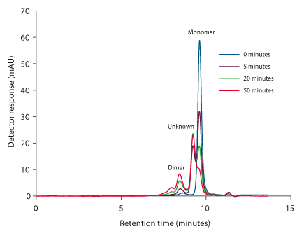
Figure 1 shows the use of a TSKgel UltraSW Aggregate column for the analysis of mAb aggregates formed by the forced acid denaturation of a monoclonal antibody. After reducing the pH of the mAb-02 sample solution to 4.7 by dilute phosphoric acid, aliquots were analyzed at 5, 20 and 50 minutes and the response was compared to that of the original sample solution.
The degradation of the monoclonal antibody creates a larger MW entity (unknown) that elutes directly after the dimer and before the monomer. Continued decay increases both peaks, but more so for the dimer. Clearly, the dimer peak height increases while the peak height of the monomer decreases. Hints of higher order ‘multimers’ show between 7 and 8 minutes.
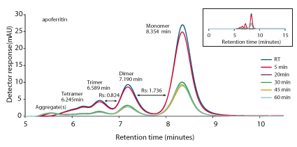
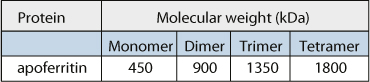
The analysis of a heat denatured, large hydrophobic metalloprotein, apoferritin, is shown in Figure 2. A set of six, 0.3 mL HPLC vials each containing 100 μL stock solution of apoferritin was used for protein thermal denaturation. Thermal denaturation was carried out at 60 °C using an electric heating block. Individual sample vials were tightly capped and exposed to the heat for 5, 20, 30, 45, and 60 minutes. Samples were analyzed using a TSKgel UltraSW Aggregate column at the end of each incubation time period. The TSKgel Ultra SW Aggregate column yielded high resolution between the monomer and dimer. The trimer, tetramer and higher order aggregates of apo-ferritin were well separated.
Conclusions
The results of both analyses demonstrate the high resolution separation of the aggregates of a monoclonal antibody and the large metalloprotein, apoferritin, generated by forced denaturation using a TSKgel UltraSW Aggregate column. With the increasing use of monoclonal antibodies in biotherapeutics, the TSKgel UltraSW Aggregate offers superior analysis of aggregates.
The TSKgel UltraSW Aggregate, 3 µm, SEC column with 30 nm pore size is specially designed with controlled pore technology which produces a shallow calibration curve in the molecular weight region of a typical monoclonal antibody. The larger pore size with an estimated exclusion limit of ~2 × 10⁶ Da provides improved resolution and quantitation of mAb aggregates and oligomers. The TSKgel UltraSW Aggregate is an excellent choice for the analysis of monoclonal antibody protein aggregates, present in their native state or when induced by forced denaturation.

TOSOH
Application n°A15L09A
Introduction
Degradation studies of biotherapeutic proteins are necessary to test their stability. The best way to test the suitability of a method is the use of real-time stability samples containing all relevant degradation products that might occur over time. Certain factors, such as product development timeline, process characteristics, excipients, and other environmental factors, however, make the use of a forced degradation study necessary. The biological phenomenon of protein aggregation is a major issue in therapeutic protein development, since the presence of these impurities reduces the potency of the drug formulation, even if non-toxic. Monoclonal antibody proteins, widely being used in the field of biotherapeutics, with the potential to replace small molecules in the future, must be free from these aggregate impurities.
In order to fully evaluate the aggregates, a size exclusion column is needed which has a large enough molecular exclusion limit, so that the higher order aggregates are not excluded in the void but separated as a function of hydrodynamic volume. This application note will show the superior resolving power of the TSKgel UltraSW Aggregate column for the analysis of monoclonal antibody and metalloprotein aggregates formed under forced denatured conditions, including acid and thermal denaturation.
Experimental Conditions
Figure 1
Column: TSKgel UltraSW Aggregate, 3 μm, 30 nm, 7.8 mm ID × 30 cm L
Mobile phase: 100 mmol/L potassium phosphate buffer, 100 mmol/L sodium sulfate, pH 6.7+0.05% NaN3
Flow rate: 1.0 mL/min
Detection: UV @ 280 nm
Temperature: ambient
Injection vol.: 10 μL
Sample: mAb-02
Concentr.: 4.5 g/L in glycine/Na phosphate, pH 6.0
Figure 2
Column: TSKgel UltraSW Aggregate, 3 μm, 30 nm, 7.8 mm ID × 30 cm L
Mobile phase: 50 mmol/L potassium phosphate (monobasic), 50 mmol/L sodium phosphate (dibasic),
100 mmol/L sodium sulfate, 0.05% NaN3, pH 6.7
Flow rate: 1.0 mL/min
Detection: UV @ 280 nm
Temperature: 30 °C
Injection vol.: 10 μL
Sample: Apoferritin Sigma, 5.0 mg/mL in 50% glycerol and 0.075 mol/L sodium chloride, stored at -20 °C
Results and Discussion
Figure 1 shows the use of a TSKgel UltraSW Aggregate column for the analysis of mAb aggregates formed by the forced acid denaturation of a monoclonal antibody. After reducing the pH of the mAb-02 sample solution to 4.7 by dilute phosphoric acid, aliquots were analyzed at 5, 20 and 50 minutes and the response was compared to that of the original sample solution.
The degradation of the monoclonal antibody creates a larger MW entity (unknown) that elutes directly after the dimer and before the monomer. Continued decay increases both peaks, but more so for the dimer. Clearly, the dimer peak height increases while the peak height of the monomer decreases. Hints of higher order ‘multimers’ show between 7 and 8 minutes.
The analysis of a heat denatured, large hydrophobic metalloprotein, apoferritin, is shown in Figure 2. A set of six, 0.3 mL HPLC vials each containing 100 μL stock solution of apoferritin was used for protein thermal denaturation. Thermal denaturation was carried out at 60 °C using an electric heating block. Individual sample vials were tightly capped and exposed to the heat for 5, 20, 30, 45, and 60 minutes. Samples were analyzed using a TSKgel UltraSW Aggregate column at the end of each incubation time period. The TSKgel Ultra SW Aggregate column yielded high resolution between the monomer and dimer. The trimer, tetramer and higher order aggregates of apo-ferritin were well separated.
Conclusions
The results of both analyses demonstrate the high resolution separation of the aggregates of a monoclonal antibody and the large metalloprotein, apoferritin, generated by forced denaturation using a TSKgel UltraSW Aggregate column. With the increasing use of monoclonal antibodies in biotherapeutics, the TSKgel UltraSW Aggregate offers superior analysis of aggregates.
The TSKgel UltraSW Aggregate, 3 µm, SEC column with 30 nm pore size is specially designed with controlled pore technology which produces a shallow calibration curve in the molecular weight region of a typical monoclonal antibody. The larger pore size with an estimated exclusion limit of ~2 × 10⁶ Da provides improved resolution and quantitation of mAb aggregates and oligomers. The TSKgel UltraSW Aggregate is an excellent choice for the analysis of monoclonal antibody protein aggregates, present in their native state or when induced by forced denaturation.
 Download_Application_a15l09a_Interchim1703.pdf
- (1.72 MiB) Downloaded 329 times